Wednesday 21 June 2023

The Anatomy of the Brainstem


Anatomy


Outside the meninges, the brainstem is shielded by the lower part of the skull. Cerebrospinal fluid (CSF) flows between the meninges and the brainstem, providing nourishment and protection.

Structure

From top to bottom, the brainstem includes the midbrain, the pons, and the medulla. Each of these sections contains nerve pathways, many of which travel throughout the whole brainstem. Cranial nerve roots are located in the brainstem, and each pair of the 12 cranial nerves emerge from the brainstem.


The cranial nerve levels are:
Cerebrum: Cranial nerves one and two
Midbrain: Cranial nerves three and four
Pons: Cranial nerves five through eight
Medulla: Cranial nerves nine through 12


The deeper portion of the brainstem is composed of grey matter, and the remaining nerve pathways of the brainstem are primarily are composed of white matter, which is more heavily myelinated (protected by a type of fat that insulates nerves).

The brainstem receives blood supply from several arteries, including the vertebral arteries, basilar artery, and pontine arteries.

Location

Located towards the back of the neck, the brainstem is the lower part of the brain, and it is continuous with the spinal cord. Behind the brainstem, the cerebellum (the part of the brain largely responsible for coordination) is also protected by the lower portion of the skull.

Anatomical Variations

The most common variations of the brainstem generally involve asymmetry of the blood supply or of the cranial nerves. These variations are usually minor, and they typically don’t cause clinical effects.


Aneurysms, which are defects in a blood vessel, can be congenital, and can develop in the blood vessels near the brainstem. Brain aneurysms near the brainstem may cause serious effects due to compression or bleeding.


Function

The brainstem contains nerves and tracts (nerve pathways) that provide motor and sensory functions throughout the body. Nerve tracts are composed of a sequence of nerves that rapidly send messages along a specific route.


Major nerve pathways in the brainstem include:
Spinothalamic: This tract runs at the outer portion of the brainstem, relaying messages of sensation that originate in sensory nerves to the spinal cord, through the brainstem, and to the thalamus in the cerebral cortex.
Corticospinal: This tract runs medially, near the center of the brainstem, sending messages from the motor portion of the cerebral cortex through the brainstem, to the spinal cord, and eventually to the muscles to control movement.
Spinocerebellar: This tract runs in the lateral portion of the brainstem, relaying messages between the cerebellum and the spinal cord to regulate the body’s position.


Some of the structures located in the brainstem work by coordinating with neurotransmitters (chemical messengers) and structures in other parts of the brain and throughout the body to control complex functions.


#Brainstem#Neuroscience#Neurology#BrainFunctions#CentralNervousSystem#NeuralPathways#BrainAnatomy#BrainHealth#NeurologicalDisorders#BrainstemFunction#BrainstemResearch#BrainstemInjury#BrainstemDysfunction#BrainstemDisorders#Neurobiology#Neuroanatomy#BrainstemAwareness#BrainstemNeurons#BrainstemHealth#BrainstemDevelopment


Friday 16 June 2023

3D printing of brain blood vessels could revolutionise neurosurgery–new technique shows how


A new 3D-printing technique using silicone can make accurate models of the blood vessels in your brain, enabling neurosurgeons to train with more realistic simulations before they operate, according to our recently published research.

Many neurosurgeons practice each surgery before they get into the operating room based on models of what they know about the patient’s brain. But the current models neurosurgeons use for training don’t mimic real blood vessels well. They provide unrealistic tactile feedback, lack small but important structural details and often exclude entire anatomical components that determine how each procedure will be performed. Realistic and personalized replicas of patient brains during pre-surgery simulations could reduce error in real surgical procedures.


3D printing, however, could make replicas with the soft feel and the structural accuracy surgeons need.

3D printing is typically thought of as a process that involves laying down layer after layer of melted plastic that solidifies as a self-supporting structure is built. Unfortunately, many soft materials do not melt and re-solidify the way the plastic filament that 3D printers typically employ do. Users only get one shot with soft materials like silicone – they have to be printed while in a liquid state and then irreversibly solidified.


3D-printing silicone with AMULIT

As researchers working at the interface of soft matter physics, mechanical engineering and materials science, we decided to tackle the problem of interfacial tension by developing a support material made from silicone oil.


#3DPrinting#BrainBloodVessels#NeurovascularPrinting#Medical3DPrinting#VascularModeling#BrainAnatomy#MedicalImaging#Cerebrovascular#Neurology#Neurosurgery#HealthTech#MedicalInnovation#PrecisionMedicine#Bioengineering#Neuroscience#PatientCare#MedicalVisualization#InnovationInMedicine#Bioprinting#ResearchAndDevelopment


Thursday 15 June 2023

The Incredible Complexity of the Cerebral Cortex Revealed

Suprascapular Nerve Block

What is the Suprascapular nerve block?


What are the Indications of the Suprascapular Nerve Block?

A suprascapular nerve block may be recommended for the treatment of:Acute pain from a shoulder injury
  • Pain following shoulder surgery
  • Adhesive capsulitis also called frozen shoulder
  • Rotator cuff injuries
  • Arthritis
  • Bursitisand cancer pain
  • Prior to the Procedure

Before administering a suprascapular nerve block, your general health is reviewed. You should inform your doctor about any medical conditions or allergies as well as recent or current medications you are taking. You are advised to stop any blood thinning medication before your procedure.


How is the Suprascapular Nerve Block Procedure performed?

For the suprascapular nerve block, you will be seated or lying on your side. Sedation may be administered to keep you comfortable. The area over your shoulder blade is cleaned using an antiseptic solution. Your doctor will locate the suprascapular nerve using your anatomy. The suprascapular nerve passes through a bony notch at the top of the shoulder blade and is targeted at this region. A nerve stimulating needle is inserted to help locate the nerve. You will usually experience a tingling sensation or muscle twitch if the needle is inserted at the right spot. Ultrasound imaging may also be used to locate the suprascapular nerve. Once the nerve is located, local anesthetic with steroid medication is carefully injected via a needle. The needle is then withdrawn, and a band-aid is placed over the site. The procedure usually takes around 10 to 15 minutes.

Postoperative Care for the Suprascapular Nerve Block Procedure

Following the procedure, your shoulder will feel numb for a short time from the local anesthetic. The pain may return once the anesthetic wears off and you may feel soreness at the injection site.

The steroid can take up to 2 weeks to begin working and provide pain relief that can last a few weeks to a few months. Sometimes the medication does not provide any relief at all and will depend on the person.Avoid driving yourself home if you have received sedation for the procedure.
Avoid straining the shoulder for at least 24 hours after your procedure.
Any swelling at the site of injection may be treated by applying ice to the area.

What are the Risks and Complications of Suprascapular Nerve Block?

Although suprascapular nerve block is a relatively safe procedure, it may be associated with complications such as:Bleeding or infection at the injection site
  • Nerve injury
  • Pneumothorax
  • Allergic reaction

#SuprascapularNerveBlock#PainManagement#ShoulderPainRelief#NerveBlockProcedure#MedicalIntervention#Anesthesia#Orthopedics#MusculoskeletalPain#ChronicPain#InterventionalPainManagement#NerveBlockTherapy#ShoulderPainTreatment#MinimallyInvasiveProcedure#NerveBlockInjection#PainReliefOptions#NonSurgicalTreatment#PainFreeLiving#MedicalProcedure#PatientCare#PhysicalTherapy

Tuesday 13 June 2023

The Power of Machine Learning in Cognitive Computing

Need a mental health day but worried about admitting it? You’re not alone

 There are days when it’s hard to face work, even when you aren’t physically sick. Should you take a day off for your mental health? If you do, should you be honest about it when informing your manager?

If you work for an organisation or in a team where you feel safe to discuss mental health challenges, you are fortunate.

Despite all the progress made in understanding and talking about mental health, stigma and prejudices are still prevalent enough to prevent many of us from willingly letting bosses and coworkers know when we are struggling.

Mental health challenges come in different forms. For some it will be a severe lifelong struggle. For many others the challenge will be periods of feeling overwhelmed by stress and needing a break.


Globally, the World Health Organisation estimates about 970 million people – about one in eight people – is suffering a mental disorder at any time, with anxiety-related disorders affecting about 380 million and depression about 360 million.

These numbers have jumped about 25% since 2019, a rise credited to the social isolation, economic hardship, health concerns and relationship strains associated with the pandemic.

But declining mental health is a longer-term trend, and it’s likely work demands have also played a role. Research identifies three main workplace contributors to mental ill-health: imbalanced job design when people have high job demand yet low job control, occupational uncertainty, and lack of value and respect.

This at least partly explains why depression and anxiety appear to be more prevalent in wealthy industrialised nations. In the United States, for example, it is estimated more than half of the population will experience a diagnosable mental disorder at some point during their lifetime.


Visit: neurology.pencis.com


#MentalHealth#MentalWellness#MentalHealthMatters#EndTheStigma#BreakTheStigma#MentalHealthAwareness#MentalHealthSupport#MentalHealthWarrior#SelfCare#SelfLove#SelfCompassion#MentalHealthAdvocate#YouAreNotAlone#Mindfulness#PositiveMentalHealth#MentalHealthRecovery#AnxietyAwareness#DepressionAwareness#BipolarDisorder#PTSDAwareness#SuicidePrevention#EndSuicide#MentalHealthFirstAid#Wellbeing#TherapyWorks#MentalHealthResources#MentalHealthEducation#MentalHealthMovement#EmotionalWellness#MindOverMatter


Monday 12 June 2023

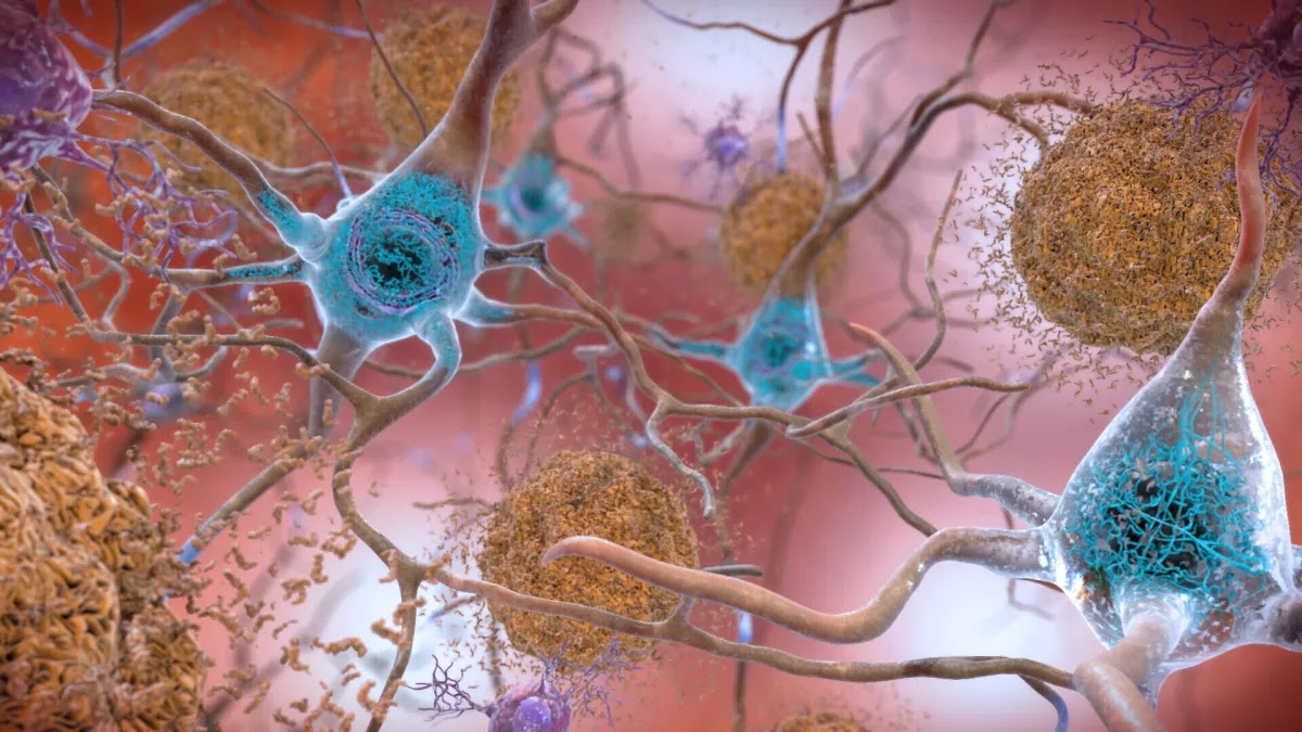
Opinion: New Alzheimer’s drugs are costly and controversial. Are we going about this all wrong?


The drugs come at a high cost for relatively limited benefits, offering only a brief respite in the pace of cognitive decline. This reality raises a question: Are we fixated on such marginally effective treatments at the expense of more promising preventive measures?

The pharmaceutical industry stands to profit substantially from these drugs, which promise to bring in billions. Eli Lilly’s stock price jumped 6.7% after it announced positive results from a trial of one new Alzheimer’s drug, increasing the company’s value by more than $25 billion that day. This isn’t surprising given that Alzheimer’s disease is estimated to be a $500-billion problem today and, due to our aging population, projected to double in cost by 2050.

Despite the market and media frenzy around the new drugs, their efficacy is contested. When another Alzheimer’s drug, aducanumab, was approved in 2021, three scientists resigned from an FDA advisory committee over the lack of evidence of its efficacy. The role of the drugs’ main target, the amyloid plaques associated with the disease, continues to be disputed.

Beneath the facade of these pharmaceutical victories, however, the potential of prevention remains vastly untapped and underreported.

The prevailing Alzheimer’s narrative, perpetuated by the dearth of effective treatments, is of inevitability: The disease is seen as an unstoppable force. This perception overshadows preventive strategies that are not only affordable but effective. Prevention can offer years of cognitive health, not just fleeting months of slowed decline.

Humpty Dumpty is an apt analogy here: It’s easier to prevent his great fall than to put him together again after he’s broken. Similarly, preserving brain cells while they are healthy is far more feasible than attempting to regenerate them after they and their myriad interconnections have been lost. Indeed, these two approaches — prevention versus late-stage reversal — don’t even fall within the same realm of difficulty.

The bias against prevention reflects a larger problem within our biomedical-industrial complex, which predominantly manages disease instead of promoting health. The American healthcare system, which consumes nearly a fifth of all expenditures, is reactive and lucrative, rushing to patch up problems after they arise rather than actively addressing risks. It’s designed to highly compensate years of sick life but not healthy life.

We champion a healthcare system focused on extending health over responding to disease. In addressing Alzheimer’s, this translates into prioritizing prevention strategies that can preserve cognitive health at a fraction of the cost of late-stage drug interventions.

But what does prevention entail? There are several often-overlooked precautions that can play a crucial role in preventing dementias such as Alzheimer’s.

These include maintaining brain oxygenation, ensuring sufficient intake of certain nutrients, managing stress, limiting inflammation, minimizing diabetes, avoiding head injuries, reducing air pollution exposure, treating hearing impairments, maintaining a healthy weight, keeping our minds active and fostering robust social connections.


Lifestyle modifications like quitting smoking and moderating alcohol consumption can also help significantly. So can reducing hypertension through medication or lifestyle changes. Eating a balanced diet, getting enough sleep and avoiding chronic infections can keep inflammation under control.

Though the impact of each of these practical, affordable measures might be modest on its own, they can work together to delay cognitive decline for years.

The effectiveness of prevention-based strategies has been shown in studies such as the Finnish Geriatric Intervention Study to Prevent Cognitive Impairment and Disability, commonly known as FINGER. The landmark trial involving more than 1,200 people has significantly outperformed any drug trial to date by tackling dementia prevention from multiple angles, ranging from nutrition and physical activity to mental and social stimulation.

After two years, the group assigned to the prevention strategies did 25% better than the control group on an assessment of overall cognitive performance. Specifically, participants showed improvements in executive functioning and processing speed, key cognitive functions often affected early in Alzheimer’s disease. They also experienced broader health benefits; the side effects of prevention are often positive rather than negative.

The success of the FINGER trial has inspired a new wave of research, with several countries now conducting their own versions, including the U.S. Study to Protect Brain Health Through Lifestyle Intervention to Reduce Risk, or POINTER. The global scientific community is recognizing the value of prevention.

The fight against Alzheimer’s is at a critical juncture. Prevention and treatment should be seen not as mutually exclusive but rather as facets of a comprehensive approach to this formidable disease. But the emphasis we place on each path has far-reaching implications not only for the millions directly affected but also for the trajectory of our healthcare system.

While it remains crucial to continue developing innovative treatments, we have at our disposal a range of accessible and cost-effective preventive measures bolstered by scientific research. These measures will not drain our pockets but will require us to recalibrate our lifestyles and priorities.

Individuals as well as healthcare providers, policymakers and society need to redirect more of our focus and resources to Alzheimer’s prevention. This could save billions in healthcare costs and, more important, preserve the memories and identities of countless individuals for longer.

In a world where Alzheimer’s is a growing threat, we can write a different narrative in which the disease is less of an inevitability and more of a challenge, one we have the tools to combat. Let’s ensure that we have as few afflicted loved ones as possible.

Nathan Price is the chief scientific officer of Thorne HealthTech and a professor on leave from the Institute for Systems Biology. Leroy Hood is the chief executive of Phenome Health and a co-founder and professor at the Institute for Systems Biology. They are the authors of “The Age of Scientific Wellness.”


#Alzheimers#EndAlzheimers#MemoryLoss#BrainHealth#Dementia#CaregiverSupport#ForgetMeNot#ResearchForACure#AwarenessMatters#RememberingLovedOnes#AlzheimersResearch#MentalHealth#CognitiveDecline#BrainAwareness#MemoryCare#SupportingFamilies#AlzheimersPrevention#Mindfulness#EndStigma#DontForgetMe

Friday 9 June 2023

5th Edition of International Research Awards on Neurology and Neuro Diso...

Research sheds new light on the genetic architecture of bipolar disorder


Bipolar disorder (BD) is a major psychiatric condition that afflicts about 1% of people. Symptoms of BD include sudden onset of depressive mood with loss of interest which alternates with a manic state of hyperactivity. The suffering of the patients and societal cost of this disorder requires the use of continued therapeutic management. Current medications-; although vital for patients with BD-;are not perfect solutions, given their potential side-effects and treatment resistance. This necessitates the development of better therapeutics for BD, including precision medicine. A major hindrance to this process, however, lies in our limited understanding of the underlying biological mechanisms of BD, i.e., its pathogenesis and the genetic architecture of people with BD. Several studies have linked BD with hereditary mutations, but recent genomic studies are now focusing on somatic mosaic variants-; new mutations occurring during developmental stages-;as another possible mechanism behind psychiatric disorders like BD.

In a new study published in Molecular Psychiatry on May 30, 2023, a team of researchers led by Associate Professor Masaki Nishioka of Juntendo University, Japan, investigated the association between mosaic variants and the risk of BD. The research team included Dr. Tadafumi Kato, also from Juntendo University, and Dr. Atsushi Takata from RIKEN Center for Brain Science. "Most analyses exploring the genetic mechanisms of BD involve extracting information from mutations that are shared among all the cells of the patients. However, mosaic de novo mutations or somatic mutations, which arise during development, are not shared among all the cells. We know very little about how these mutations influence diseases like BD. Therefore, for our study, we hypothesized that deleterious mosaic de novo variants (mDNVs) in the genes associated with developmental disorders may have a role in BD's pathology," explains Dr. Nishioka.

The team recruited 235 participants with BD and 39 control participants without psychiatric disorders. They collected blood or saliva samples from the participants and analyzed the DNA extracted from these samples using deep exome sequencing (DES) to detect mosaic variants that originated during early development. Participants with BD had mosaic variants enriched in genes that are responsible for causing developmental disorders (DD) and autism spectrum disorder (ASD). Moreover, the proteins encoded by the DD/ASD genes with the proteins of the mosaic variants were closely linked and had more protein-protein interactions than expected.

Surprisingly, the team also found significant heteroplasmic mutations (another class of mosaic variants) in mitochondrial tRNA genes of participants with BD. For reference, some tRNA mutations are known to be pathogenic for other diseases. In fact, two participants with mitochondrial tRNA mutations had recurrent m.3243 A > G variants, which are known to be major causal variants for mitochondrial diseases, MELAS, which is a serious neurodevelopmental disorder. This finding complements other studies that have found that patients with mitochondrial diseases often exhibit symptoms of bipolar disorder or schizophrenia.

Furthermore, both the sets of deleterious mosaic variants-;mDNVs and mitochondrial tRNA variants-;were either absent or rarely observed in the control participants. These results indicate that the molecular mechanisms underlying DD/ASD could also contribute to BD in a compromised way through mosaic mutations. Moreover, they suggest that mitochondrial tRNA variants could be associated with BD despite the patient showing no obvious symptoms of mitochondrial diseases.

With this study, the researchers demonstrate that mosaic mutations, particularly those in neurodevelopmental disorder genes and mitochondrial tRNA genes, may be involved in the pathophysiology of BD. Dr. Nishioka is encouraged by what their study's findings mean for scientists pursuing the research of molecular pathologies in neuropsychiatric diseases. He concludes, "Our research sheds new light on the genetic architecture of BD and provides more insights into the pathological contribution of mosaic variants in human diseases. This could potentially pave the way and expedite new research for the development of more effective, precision medications for treating BD and other psychiatric disorders."


Visit: neurology.pencis.com


#MentalHealth#Depression#Anxiety#BipolarDisorder#MoodSwings#MentalIllness#MentalHealthAwareness#SelfCare#Wellbeing#EndTheStigma#YouAreNotAlone#MentalHealthMatters#Recovery#Therapy#Mindfulness#MoodDisorderAwareness#MentalHealthSupport#HealthyMind#EmotionalWellness

Saturday 3 June 2023

Pediatric Neurology 101

Pediatric Neurology Devices Market expected to reach a value of USD 3.26 billion by 2029. The market is anticipated to grow at a CAGR of 6.3% during the forecast period.


The research methodology employed for this report includes extensive primary and secondary research. The revenue impact of the COVID-19 pandemic on market leaders, followers, and disrupters has been analyzed and reflected in the report's findings.

What are Pediatric Neurology Devices Market Dynamics?

The report identifies several factors that contribute to the growth of the pediatric neurology devices market, including the adoption of advanced technology by developing economies, government support for research and development, and the launch of medical devices for treating neurological disorders in children. However, the high cost associated with diagnosing and monitoring neurological diseases may hinder market growth.

What are Pediatric Neurology Devices Market regional insights:


North America holds the largest market share in the pediatric neurology devices market, driven by the rising incidence of neurological diseases such as depression, epilepsy, and migraine. The region benefits from high healthcare spending and government support for research and development. Additionally, international conferences and events like the 3rd International Conference on Neurology and Brain Disorders held in Dublin, Ireland, contribute to regional growth.

1. Global Pediatric Neurology Devices Market Segmentation


• Global Market, by Product (2021-2029)
• Global Market, by Service (2021-2029)
• Global Market, by Application (2021-2029)
• Global Market, by Neurological Subspecialties (2021-2029)


2. Regional Pediatric Neurology Devices Market (2021-2029)


• Regional Market, by Product (2021-2029)
• Regional Market, by Service (2021-2029)
• Regional Market, by Application (2021-2029)
• Regional Market, by Neurological Subspecialties (2021-2029)
• Regional Market, by Country (2021-2029)


3. Company Profile: Key players


• Company Overview
• Financial Overview
• Global Presence
• Capacity Portfolio
• Business Strategy
• Recent Developments


#PediatricNeurology#ChildNeurology#PediatricNeurologist#Neurodevelopment#PediatricNeuroscience#PediatricBrainDisorders#ChildhoodEpilepsy#PediatricSeizures#NeurologicalDevelopment#PediatricStroke#PediatricHeadaches#NeurologicalConditions#PediatricNeuromuscularDisorders#PediatricNeuroRehab#PediatricNeuroSurgery#NeurodevelopmentalDisorders#PediatricNeurologyclinic#PediatricNeuroResearch#PediatricNeurotherapeutics#PediatricNeuroImaging